
Eyeball spots may be caused by a variety of different medical conditions or diseases. While some may be relatively harmless, others may be the result of significant health problems. Discoloration may appear as a brown, pink, red, yellow, or white spot on the eyeball, making those affected feel concerned that their vision is at risk.
Causes of eyeball spots
Everything from minor trauma to cancer can cause the development of eyeball spots. Most are non-serious and present without any additional symptoms, but it is recommended to see an optometrist if you notice any change in your eyes or vision. Eyeball spots are generally very easy to identify, as they contrast with the rest of the eye.
The visible surface of the eyeball consists of the cornea (the clear front window of the eye), the pupil (the dark aperture of the iris that focuses light rays onto the retina), the iris (colored part of the eye that regulates the amount of light that enters), the sclera (the whites of the eyes) and the conjunctiva (a highly vascularized covering of the sclera).
The following are various medical conditions that may be possible causes of eyeball spots.
Axenfeld nerve loop: Long posterior ciliary nerve loops found within the sclera. Loops may appear as grey or white nodules under the conjunctiva and are often surrounded by pigment. Axenfeld nerve loops are fairly common and may be confused as being an embedded foreign body in the eye.
Crystal deposits: The use of vitamin C eye drops can cause crystal deposits to form in the cornea of the eye.
Horner-Trantas dots: Characterized by small rounded protuberances, called papilla, found on the surface of the eye or the eyelid. Additional symptoms include severe itching, photophobia, foreign body sensation, mucous discharge, blepharospasm (abnormal contraction of the eyelid), and blurring of vision. This condition often affects young males in hot dry climates in a seasonal manner and is thought to be an allergic and inflammatory response.
Nevus: Occasionally called a “freckle of the eye,” similar to a mole found on the skin. A nevus is a colored growth in front of the eye, around the iris, or under the retina at the back of the eye. They are usually harmless, but could possibly develop into cancer of the eye.
Pinguecula: Described as being a yellowish, slightly raised thickening of the conjunctiva. They are non-cancerous and appear as bumps on the eyeball, typically found on the top of the middle part of the sclera. Pinguecula is believed to be caused by ultraviolet radiation exposure from the sun, but frequent exposure to dust and wind also appear to be causative risk factors. This condition can be seen more commonly in middle-aged or older people who spend a lot of time outdoors without sunglasses and hats to protect their eyes from harmful sun rays.
Pterygium: Also known as surfer’s eye, this condition is described as being a growth of pink, fleshy tissues on the conjunctiva. It generally forms on the side closest to the nose and can grow towards the pupil area. Despite its disturbing appearance, pterygium isn’t cancer, with its growth typically slowing down over time or stopping at a certain period. Extreme cases see a partial or complete coverage of the pupil causing vision disturbances.
Racial melanosis: A benign, bilateral condition found mostly in darkly pigmented individuals. They are characterized by flat conjunctival pigmentation or dark spots. This condition rarely develops into conjunctival melanoma.
Scleral thinning: Can occur due to various conditions such as various conditions, including myopic degeneration, chronic scleritis, local scleral pathologies, and scleral injury. It is characterized by the thinning of the white part of the eyes known as the sclera and can be due to excessive use of cautery in the scleral bed, overuse of antimetabolites, prolonged irradiation, transscleral diode laser cycloablation, strabismus surgery, and deep sclerectomy procedures.
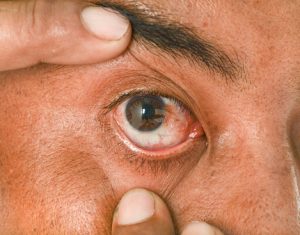
Subconjunctival hemorrhage: Appears as a red area on the sclera and is due to small vessels within the conjunctiva breaking spontaneously or due to injury. These red spots can be easily spotted and typically do not present with any additional symptoms. Subconjunctival hemorrhage tends to clear without treatment in one or two weeks.
Corneal ulcers: Known as an open sore on the cornea and has a wide variety of causes including infection, physical, and chemical trauma. Ulcers on the cornea can cause serious problems and may result in vision loss or even blindness. Most cases improve with appropriate treatment.
Conjunctival melanoma: A cancerous, life-threatening growth that appears as a brown spot on the eye. They are typically painless, flat, and have a brownish discoloration. They are often mistaken for a benign freckle on the eye but may evolve into one or more brown or pink nodules on the eye. Conjunctival melanoma is more commonly seen in Caucasian individuals.
Conjunctival squamous carcinoma: A type of cancer found on the surface of the eye more commonly seen in older Caucasian individuals. They may appear as white or yellow-pink nodules that are easily seen upon inspection. It is believed that this condition is caused by excessive exposure to sunlight.
Osteogenesis imperfecta (OI): A congenital disease frequently caused by a defect in the gene that produces type one collagen, an important component for the formation of bone, skin, and dentin. The severity of this condition depends on the specific gene defect. Symptoms include having a blue tint on the whites of the eyes, multiple bone fractures, and hearing loss. Because type one collagen affects many structures in the body, those affected may eventually develop bowed legs and arms as well.
Uveitis and iritis: Inflammation of the middle part of the eye called the uvea and consists of the iris, ciliary body, and choroid. Possible causes include eye injury and inflammatory disease. Additionally, exposure to toxic chemicals such as pesticides and acids may also cause the condition. Typically, symptoms include red eyes, eye pain, light sensitivity, and decreased visual acuity.
Complications of eyeball spots
Ultimately, the underlying cause of your eyeball spot will determine what kind of complications you will develop. However, for the most part, eyeball spots do not present with serious complications but may have an associated itchy or gritty feeling. Depending on the cause of your eyeball spot, it may cause significant discomfort, leave scarring, or even develop into cancer.
Once the underlying cause of your particular eyeball spot condition has been identified by an experienced medical professional, tailored treatment and conservative management can begin.